Novel Choline-Deficient and 0.1%-Methionine-Added High-Fat Diet Induces Burned-Out Metabolic-Dysfunction-Associated Steatohepatitis with Inflammation by Rapid Immune Cell Infiltration on Male Mice – PubMed Black Hawk Supplements
BLACK HAWK: Best lions mane supplement for stress
Published article
Background: Metabolic-dysfunction-associated steatotic liver disease (MASLD) is a progressive liver disorder that possesses metabolic dysfunction and shows steatohepatitis. Although the number of patients is globally increasing and many clinical studies have developed medicine for MASLD, most of the studies have failed due to low efficacy. One reason for this failure is the lack of appropriate animal disease models that reflect human MASLD to evaluate the potency of candidate drugs. Methods: We…
Black Hawk Supplements, best supplements in the UK

Novel Choline-Deficient and 0.1%-Methionine-Added High-Fat Diet Induces Burned-Out Metabolic-Dysfunction-Associated Steatohepatitis with Inflammation by Rapid Immune Cell Infiltration on Male Mice
Takatoshi Sakaguchi et al. Nutrients. .
Abstract
Background: Metabolic-dysfunction-associated steatotic liver disease (MASLD) is a progressive liver disorder that possesses metabolic dysfunction and shows steatohepatitis. Although the number of patients is globally increasing and many clinical studies have developed medicine for MASLD, most of the studies have failed due to low efficacy. One reason for this failure is the lack of appropriate animal disease models that reflect human MASLD to evaluate the potency of candidate drugs. Methods: We developed a novel choline-deficient and 0.11%-methionine-added high-fat diet (CDAHFD)-based (MASH) diet that can induce murine metabolic-dysfunction-associated steatohepatitis (MASH) without severe body weight loss. We performed kinetic analyses post-feeding and proposed an appropriate timing of MASH pathogenesis by quantitatively analyzing steatosis, inflammation, and fibrosis. Results: This MASH diet induced liver fibrosis earlier than the conventional CDAHFD model. In brief, lipid accumulation, inflammation, and fibrosis started after 1 week from feeding. Lipid accumulation increased until 8 weeks and declined thereafter; on the other hand, liver fibrosis showed continuous progression. Additionally, immune cells, especially myeloid cells, specifically accumulated and induced inflammation in the initiation stage of MASH. Conclusions: The novel MASH diet promotes the dynamics of lipid deposition and fibrosis in the liver, similar to human MASH pathophysiology. Furthermore, immune-cell-derived inflammation possibly contributes to the initiation of MASH pathogenesis. We propose this model can be the new pre-clinical MASH model to discover the drugs against human MASH by evaluating the interaction between parenchymal and non-parenchymal cells.
Keywords: CDAHFD; MASLD; burned-out MASH; hepatic inflammation; mitochondrial dysfunction.
Conflict of interest statement
Y.N. declares to be an employee and shareholder of Otsuka Pharmaceutical Co., Ltd. H.M. declares to be an employee of Oriental Yeast Co., Ltd. The remaining authors declare that the research was conducted in the absence of any commercial or financial relationships that could be construed as a potential conflict of interest.
Figures

Schematic diagram of experimental design.

MASH diet feeding caused hepatic steatosis, inflammation, and tumorigenesis. (A) Schematic diagram of experimental procedure; (B) body weight (n = 5–6); (C) changes in the appearance of the digestive tract over time; (D) the appearance of the tumor; hematoxylin and eosin (H,E) staining of (E) the tumor (scale bars, 500 μm (left) or 100 μm (right)), and (F) the liver, scale bars, 100 μm; (G) scores of steatosis and lobular inflammation, and NAFLD activity score (NAS) (n = 4–10); (H) serum AST and ALT levels (n = 3). Zero weeks (0W) in the figure indicates Chow-diet-fed control mice. Values are presented as mean ± S.E.M. * p < 0.05, ** p < 0.01, *** p < 0.001, and **** p < 0.0001 vs. 0-week group. Statistical significance was calculated by a one-way ANOVA with Dunnett’s post hoc test.

MASH diet feeding caused hepatic fibrosis. (A,B) Sirius red (SR) staining of the liver (n = 4–5); (C,D) the protein expression of COL1A1 and α-SMA in the liver (n = 3); (E) the mRNA expression of Col1a1, Acta2, and Tgfb1 in the liver (n = 4–9). Zero weeks (0W) in the figure indicates Chow-diet-fed control mice. Values are presented as mean ± S.E.M. * p < 0.05, ** p < 0.01, *** p < 0.001, and **** p < 0.0001 vs. 0-week group. Statistical significance was calculated by a one-way ANOVA with Dunnett’s post hoc test.

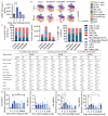
Effects of MASH diet feeding on inflammation in the early stage. (A) Total number of CD45+ cells in the liver (n = 3); (B) representative images of profiling CD45+ cells in the liver and blood by CyTOF visualized by tSNE; (C) Percentage and the number of CD45+ cells in the liver and percentage of the CD45+ cells in the blood by CyTOF (n = 3); (D) the mRNA expression of Tnf, Il6, Il1b, and Ifng in the liver (n = 3–9). Zero weeks (0W) in the figure indicates Chow-diet-fed control mice. Values are presented as mean ± S.E.M. * p < 0.05, ** p < 0.01, and *** p < 0.001 vs. 0-week group in (A,C); a p < 0.05 vs. Chow group, b p < 0.05 vs. NASH1W, and c p < 0.05 vs. NASH4W group in (B). Statistical significance was calculated by a one-way ANOVA with (A,C) Dunnett’s post hoc test; (B) Tukey’s post hoc test.

Effects of MASH diet feeding on lipid accumulation and de novo lipogenesis. (A,B) Oil Red O staining in the liver (n = 3–6); (C–F) the protein expression of AceCS1, ACL, p-ACL, ACC, p-ACC, Fas, AMPK, p-AMPK, and SREBP-1 in the liver (n = 3). Zero weeks (0W) in the figure indicates Chow-diet-fed control mice. Values are presented as mean ± S.E.M. * p < 0.05, ** p < 0.01, *** p < 0.001, and **** p < 0.0001 vs. 0-week group. Statistical significance was calculated by a one-way ANOVA with Dunnett’s post hoc test.

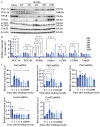
Effects of MASH diet on mitochondrial homeostasis. (A,B) The protein expression of PGC-1α, PGC-1β, PPARγ, Parkin, LC3B, and Tom20 in the liver (n = 3); (C) the mRNA expression of Pck2, Pdk4, Plin2, Cox4i1, and Cox4i2 in the liver (n = 3–9). Zero weeks (0W) in the figure indicates Chow-diet-fed control mice. Values are presented as mean ± S.E.M. * p < 0.05, ** p < 0.01, *** p < 0.001, and **** p < 0.0001 vs. 0-week group. Statistical significance was calculated by a one-way ANOVA with Dunnett’s post hoc test.
References
MeSH terms
Substances
Grants and funding
LinkOut – more resources
-
Full Text Sources
BLACK HAWK: Best shilajit supplement for men
Read the original publication: